
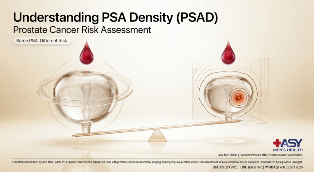
A clinical guide on calculating PSA Density, differentiating Benign Prostatic Hyperplasia (BPH) from cancer, and optimizing biopsy decisions.
Book ConsultationQuick Summary
- Volume-Adjusted Biomarker: PSA Density (PSAD) is calculated by dividing total serum PSA (ng/mL) by total prostate gland volume (cc) measured via ultrasound or MRI.
- Resolves PSA Ambiguity: A large prostate gland affected by Benign Prostatic Hyperplasia (BPH) naturally produces more PSA. PSAD distinguishes whether elevated PSA is due to gland size or malignant cell activity.
- Clinical Cut-Off (0.15 ng/mL/cc): A PSAD value greater than 0.15 ng/mL/cc indicates a significantly higher risk of clinically significant prostate cancer.
- Reduces Unnecessary Biopsies: Combined with mpMRI (PI-RADS scoring), PSAD helps up to 30–40% of men safely avoid invasive prostate biopsies.
- Guides Biopsy Decisions in PI-RADS 3: Crucial for borderline cases (PI-RADS 3), determining whether active monitoring or targeted fusion biopsy is required.
Table of Contents
1. What Is PSA Density (PSAD)? 2. How PSA Density Is Calculated 3. Differentiating BPH from Prostate Cancer 4. Clinical Thresholds & Risk Stratification 5. Integrating PSAD with mpMRI & PI-RADS 6. The ASY Clinic Urological Health Protocol 7. Key Takeaways1. What Is PSA Density (PSAD)?
Prostate-Specific Antigen (PSA) is a protein produced by both normal and malignant prostate glandular cells. While serum PSA testing is the standard screening tool for prostate disease, elevated PSA alone is not specific to cancer—conditions such as Benign Prostatic Hyperplasia (BPH), prostatitis, or trauma also elevate PSA levels.
PSA Density (PSAD) addresses this limitation by measuring the amount of PSA produced per unit volume of prostate tissue. Because cancer cells release significantly more PSA into the bloodstream per unit volume than benign enlarged tissue, PSAD serves as a far more accurate secondary biomarker for assessing malignancy risk.
2. How PSA Density Is Calculated
The calculation of PSA Density requires two distinct clinical data points:
Formula:
PSA Density (ng/mL/cc) = Total Serum PSA (ng/mL) ÷ Prostate Volume (cc)
- Total Serum PSA: Obtained via a standard venous blood test.
- Prostate Volume: Accurately measured in cubic centimeters (cc or mL) using Transrectal Ultrasound (TRUS) or 3T Multiparametric MRI (mpMRI) using the prolate ellipsoid volume formula:
Length × Width × Height × 0.52.
3. Differentiating BPH from Prostate Cancer
Understanding why PSAD is vital requires examining how prostate volume influences serum PSA levels:
| Clinical Scenario | Prostate Volume | Total PSA | Calculated PSAD | Clinical Interpretation |
|---|---|---|---|---|
| Patient A (Severe BPH) | 80 cc | 6.5 ng/mL | 0.08 ng/mL/cc | Low Risk (Elevated PSA is due to large benign gland size). Biopsy usually avoided. |
| Patient B (Early Malignancy) | 25 cc | 6.5 ng/mL | 0.26 ng/mL/cc | High Risk (Elevated PSA relative to a small gland). Targeted biopsy recommended. |
As shown above, both patients have an identical Total PSA of 6.5 ng/mL. Without PSAD, both might be advised to undergo an invasive biopsy. However, calculating PSAD reveals that Patient A simply has a large benign gland, whereas Patient B harbors a high risk of active cancer.
💡 Clinical Pearl: The BPH Volume Rule
On average, benign prostate tissue (BPH) increases total serum PSA by approximately 0.1 ng/mL per gram (or cc) of tissue. If serum PSA rises far above what expected gland volume accounts for, the likelihood of malignant activity increases substantially.
4. Clinical Thresholds & Risk Stratification
Urological guidelines employ specific PSAD cut-offs to guide diagnostic pathways:
- PSAD < 0.10 ng/mL/cc: Very low risk of clinically significant cancer. Reassuring finding; routine clinical monitoring advised.
- PSAD 0.10 – 0.15 ng/mL/cc: Intermediate / Borderline risk zone. Requires evaluation alongside free-to-total PSA ratio and clinical history.
- PSAD > 0.15 ng/mL/cc: High risk threshold. Strongly associated with clinically significant prostate cancer (Gleason ≥ 7). Targeted biopsy is generally indicated.
5. Integrating PSAD with mpMRI & PI-RADS
In modern precision urology, PSA Density is rarely used in isolation; it is integrated directly with Multiparametric MRI (mpMRI) and PI-RADS scoring:
- PI-RADS 1 & 2 + Low PSAD (<0.15): Confirms low risk; patients can safely defer prostate biopsy and continue PSA surveillance.
- PI-RADS 3 (Equivocal MRI): PSAD acts as the tie-breaker. A PSAD <0.15 supports active monitoring, while a PSAD >0.15 indicates the need for an MRI-TRUS Fusion Biopsy.
- PI-RADS 4 & 5: High risk regardless of PSAD, indicating the need for targeted 3D fusion biopsy.
6. The ASY Clinic Urological Health Protocol
At ASY Clinic, our urology practice uses advanced biomarker integration to prevent unnecessary invasive procedures:
- Accurate Volume Measurement: Utilizing high-resolution 3T mpMRI imaging to measure exact prostate dimensions and volume.
- Multi-Biomarker Risk Stratification: Combining Total PSA, Free PSA ratio, PSAD, and PI-RADS scores for diagnostic decisions.
- MRI-TRUS Fusion Biopsy Pathways: Direct referral for 3D targeted tissue sampling when high PSAD or elevated PI-RADS scores are confirmed.
7. Key Takeaways
- Volume-Corrected Accuracy: PSA Density adjusts raw PSA values according to prostate gland volume.
- Prevents Over-Biopsy: Helps men with benign enlarged prostates (BPH) safely avoid unnecessary needle biopsies.
- Critical 0.15 Cut-Off: A PSAD greater than 0.15 ng/mL/cc indicates an increased likelihood of significant prostate cancer.
- Decision Support for PI-RADS 3: Serves as a vital decision tool for borderline MRI findings.
Frequently Asked Questions
Why is PSA Density better than total serum PSA alone?
Total PSA can be elevated by benign conditions like Benign Prostatic Hyperplasia (BPH) or inflammation. PSA Density adjusts the serum PSA value for the overall volume of the prostate, preventing men with large benign prostates from undergoing unnecessary biopsies.
How is prostate volume measured to calculate PSAD?
Prostate volume is accurately measured using Transrectal Ultrasound (TRUS) or Multiparametric MRI (mpMRI) by calculating the dimensions (length, width, and height) of the gland in cubic centimeters (cc).
What is the clinical cut-off threshold for PSA Density?
A PSA Density cut-off of 0.15 ng/mL/cc is widely used in urology. A PSAD above 0.15 indicates a significantly higher risk of clinically significant prostate cancer, often prompting further diagnostic evaluation or biopsy.
About the Author
Dr. Sirirat Nakdang
นพ. สิริรัฐ นาคแดง
ศัลยแพทย์ทางเดินปัสสาวะและระบบสืบพันธุ์เพศชาย
Urologist & Men's Health Specialist
Sirirat Nakdang, MD
Specialist in Precision Urology, Prostate Cancer Risk Stratification & Men's Health
Thai Medical License No. ว55466
References
- Catalona, W. J., et al. (1994). Comparison of digital rectal examination and serum prostate-specific antigen in the early detection of prostate cancer. The Journal of Urology, 151(5), 1283–1290.
- Nordström, T., et al. (2018). Prostate-specific antigen density in the diagnostic pathway of prostate cancer. European Urology Focus, 4(6), 857–863.
- Mottet, N., et al. (2021). EAU Guidelines on Prostate Cancer. European Urology, 79(2), 243–262.
- Turkbey, B., et al. (2019). Prostate Imaging Reporting and Data System Version 2.1 (PI-RADS v2.1). European Urology, 76(3), 340–351.
Consultation & Prostate Risk Evaluation
Calculate your PSA Density and receive expert prostate risk stratification at ASY Clinic.
Book Consultation
Painless hematuria
Painless blood in urine (Hematuria) may appear only once and disappear, but it should never be ignored. Learn about possible causes—including stones, prostate conditions, kidney disease and urinary tract cancer—and when to see a urologist.

Peptides and Healthy Aging: Evidence vs Experimental | Peptide Pattaya
Peptides are not automatically experimental. Insulin, GLP-1 medicines, teriparatide and many other peptide drugs are established medical therapies. But many peptides promoted for longevity or anti-aging still lack robust human evidence.

STI Testing After a New Sexual Partner: What to Test & When | Pattaya
STI testing after a new sexual partner is not simply one “full panel” done on one day. Different infections require different samples and have different window periods. Learn what to test, when to test, and when repeat testing may be needed.