

A clinical analysis comparing traditional systematic prostate biopsy with 3D MRI-TRUS Fusion Biopsy for accurate cancer detection and safety.
Book ConsultationQuick Summary
- From Blind Sampling to Precision Targeting: Traditional TRUS biopsy samples prostate tissue systematically (blindly), missing up to 30% of significant cancers. MRI-TRUS Fusion Biopsy combines high-resolution 3T mpMRI scans with real-time ultrasound to target exact tumors.
- Higher Detection of Aggressive Cancers: Fusion biopsy significantly improves detection rates of clinically significant prostate cancer (Gleason score ≥ 7), ensuring timely and life-saving intervention.
- Reduces Over-Diagnosis & Excess Samples: By accurately targeting suspected lesions (PI-RADS 3–5), urologists take fewer unnecessary cores, reducing tissue trauma.
- Reaches Hidden Anterior Tumors: Traditional needle biopsies frequently miss tumors in the anterior zone or apex of the prostate; Fusion Biopsy easily navigates to these challenging anatomical sites.
- Near-Zero Sepsis with Transperineal Approach: Combining Fusion Biopsy with Transperineal insertion bypasses the rectum entirely, eliminating major post-biopsy infection risks.
Table of Contents
1. The Evolution of Prostate Tissue Sampling 2. How MRI-TRUS Fusion Biopsy Works 3. Head-to-Head Comparison: Traditional vs. Fusion Biopsy 4. Key Clinical Advantages of Fusion Biopsy 5. Transperineal Approach: Safety & Infection Prevention 6. The ASY Clinic Precision Diagnostics Protocol 7. Key Takeaways1. The Evolution of Prostate Tissue Sampling
For decades, diagnosing prostate cancer relied on Standard Systematic TRUS Biopsy (Transrectal Ultrasound-Guided Biopsy). During this procedure, a urologist inserted an ultrasound probe into the rectum and took 10 to 12 systematic needle cores randomly across standard zones of the gland.
The core limitation of standard ultrasound is that it cannot differentiate prostate cancer tissue from normal benign tissue. As a result, standard biopsy was essentially a "blind" sampling technique—frequently missing aggressive tumors while inadvertently detecting insignificant, low-grade lesions that required no treatment.
The introduction of MRI-TRUS Fusion Biopsy has transformed urology by shifting the standard of care from random sampling to image-guided precision targeting.
2. How MRI-TRUS Fusion Biopsy Works
MRI-TRUS Fusion Biopsy bridges pre-operative diagnostic imaging with intra-operative intervention in three synchronized steps:
- 3T Multiparametric MRI (mpMRI): Before the procedure, high-resolution mpMRI creates a detailed 3D map of the prostate, identifying suspicious lesions and grading them using the PI-RADS (Prostate Imaging Reporting and Data System) protocol.
- 3D Software Fusion: Advanced fusion software imports the MRI data and overlays the 3D target map directly onto live ultrasound images during the procedure.
- Targeted Needle Guidance: The urologist guides the biopsy needle directly into the mapped 3D coordinate of the lesion with millimeter precision.
3. Head-to-Head Comparison: Traditional vs. Fusion Biopsy
Evaluating both techniques side-by-side highlights why global urological guidelines (EAU and NCCN) strongly advocate for pre-biopsy MRI and targeted fusion biopsy:
| Clinical Feature | Traditional TRUS Biopsy | MRI-TRUS Fusion Biopsy |
|---|---|---|
| Targeting Method | Systematic / Blind 12-core grid | Direct 3D MRI-guided targeting |
| Significant Cancer Detection | 30–40% false-negative rate | >85–90% detection accuracy for Gleason ≥7 |
| Anterior Zone Accessibility | Poor (frequently missed) | Excellent (precise trajectory mapping) |
| Over-Diagnosis Risk | High (detects non-harmful indolent tumors) | Low (focuses strictly on aggressive lesions) |
| Infection / Sepsis Rate | 1–3% (Transrectal route) | Near 0% (Transperineal Fusion route) |
💡 Clinical Insight: The Gleason Grade Migration
Random systematic biopsies often sample the outer edge of a tumor, mistakenly misclassifying an aggressive Gleason 4+3=7 cancer as a low-grade Gleason 3+3=6. Fusion Biopsy directly targets the dense, hypercellular core of the lesion shown on MRI, yielding accurate pathological grading and preventing delayed treatment.
4. Key Clinical Advantages of Fusion Biopsy
Choosing MRI-TRUS Fusion Biopsy offers clear diagnostic benefits for patients with elevated PSA or abnormal DRE findings:
- Eliminates the "Needle in a Haystack" Problem: Rather than taking random needle passes hoping to strike a lesion, the needle is directed precisely into the suspicious node.
- Reaches Difficult Anatomical Regions: Up to 20% of prostate cancers develop in the anterior transition zone or the apex—areas virtually unreachable during standard transrectal biopsies. Fusion software recalculates needle trajectory to hit these deep targets effortlessly.
- Fewer Repeat Biopsies: Men with persistently high PSA levels who previously received "negative" blind biopsy results often find their hidden tumor accurately diagnosed on their first Fusion Biopsy.
- Sparing Healthy Tissue: Precision targeting reduces unnecessary needle puncture cores into healthy benign tissue, minimizing local swelling, bleeding, and discomfort.
5. Transperineal Approach: Safety & Infection Prevention
In addition to target accuracy, modern Fusion Biopsies are increasingly performed via the Transperineal route rather than through the rectum (Transrectal).
By passing the biopsy needle through disinfected perineal skin (between the scrotum and anus), the procedure avoids introducing rectal bacteria into the blood or prostate gland. This reduces post-biopsy sepsis and severe infection rates to virtually zero, while eliminating the need for heavy prophylactic antibiotics.
6. The ASY Clinic Precision Diagnostics Protocol
At ASY Clinic, our urological practice incorporates advanced diagnostic pathways to protect patient health and ensure accuracy:
- Pre-Biopsy Risk Stratification: Combining Total PSA, Free PSA, and PSA Density (PSAD) before recommending diagnostic imaging.
- 3T mpMRI Quality Control: Expert radiologist and urologist joint review of PI-RADS 3, 4, and 5 lesions.
- Transperineal Fusion Biopsy Pathways: Utilizing targeted 3D fusion platforms to maximize detection accuracy while prioritizing patient safety and comfort.
7. Key Takeaways
- Precision Guidance: MRI-TRUS Fusion Biopsy replaces blind systematic sampling with 3D image-guided precision.
- Detects Aggressive Cancers: Significantly increases detection rates for high-grade, clinically dangerous prostate tumors.
- Accesses Hidden Sites: Successfully samples anterior and apical zones that standard biopsies miss.
- Superior Safety Profile: When paired with a Transperineal approach, infection and post-procedure sepsis risks are virtually eliminated.
Frequently Asked Questions
How does MRI-TRUS Fusion Biopsy differ from traditional TRUS biopsy?
Traditional TRUS biopsy relies on random, systematic sampling (12-core grid) because standard ultrasound cannot distinguish cancer from normal tissue. Fusion Biopsy overlays 3T mpMRI images onto real-time ultrasound, allowing urologists to target suspicious lesions directly with millimeter precision.
Does Fusion Biopsy reduce the risk of infection or sepsis?
Yes. Fusion Biopsy is frequently performed via a Transperineal approach (through the skin between the scrotum and anus), which completely avoids rectal bacteria, lowering the infection and sepsis rate to near zero percent.
Why is targeted biopsy critical for aggressive prostate cancer?
Aggressive, high-grade tumors (Gleason score 7 or higher) often lie in anterior or difficult-to-reach zones of the prostate. Fusion biopsy accurately samples these hidden areas, preventing missed diagnoses and over-treatment of harmless low-grade tumors.
About the Author
Dr. Sirirat Nakdang
นพ. สิริรัฐ นาคแดง
ศัลยแพทย์ทางเดินปัสสาวะและระบบสืบพันธุ์เพศชาย
Urologist & Men's Health Specialist
Sirirat Nakdang, MD
Specialist in Precision Urology, Prostate Cancer Diagnostics & Fusion Biopsy
Thai Medical License No. ว55466
References
- Kasivisvanathan, V., et al. (2018). MRI-Targeted or Standard Biopsy for Prostate-Cancer Diagnosis (PRECISION Study). The New England Journal of Medicine, 378(19), 1767–1777.
- Siddiqui, M. M., et al. (2015). Comparison of MR/ultrasound fusion-guided biopsy with ultrasound-guided biopsy for the diagnosis of prostate cancer. JAMA, 313(4), 390–397.
- Mottet, N., et al. (2021). EAU Guidelines on Prostate Cancer: Update 2021. European Urology, 79(2), 243–262.
- Ahdoot, M., et al. (2020). MRI-Targeted, Systematic, or Combined Biopsy for Prostate Cancer Diagnosis. The New England Journal of Medicine, 382(10), 917–928.
Consultation & Fusion Biopsy Evaluation
Evaluate your PSA level, mpMRI scan, and suitability for targeted Fusion Biopsy at ASY Clinic.
Book Consultation
Painless hematuria
Painless blood in urine (Hematuria) may appear only once and disappear, but it should never be ignored. Learn about possible causes—including stones, prostate conditions, kidney disease and urinary tract cancer—and when to see a urologist.

Peptides and Healthy Aging: Evidence vs Experimental | Peptide Pattaya
Peptides are not automatically experimental. Insulin, GLP-1 medicines, teriparatide and many other peptide drugs are established medical therapies. But many peptides promoted for longevity or anti-aging still lack robust human evidence.
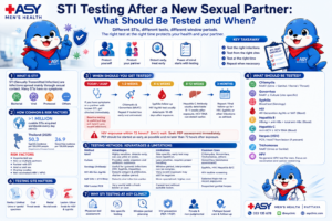
STI Testing After a New Sexual Partner: What to Test & When | Pattaya
STI testing after a new sexual partner is not simply one “full panel” done on one day. Different infections require different samples and have different window periods. Learn what to test, when to test, and when repeat testing may be needed.