
ASY Clinic · Urology · PattayaASY Clinic · คลินิกทางเดินปัสสาวะ · พัทยา
Kidney Stone Flank Pain & Renal Colicปวดสีข้างจากนิ่วในไตหรือนิ่วในท่อไต
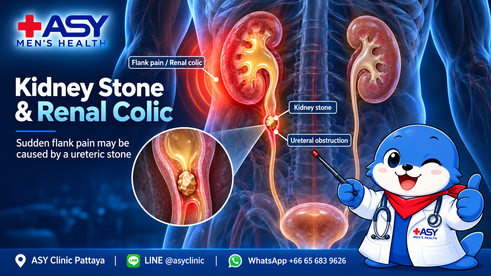
Sudden severe pain in the side or back that travels toward the lower abdomen or groin may be renal colic from a ureteric stone—but not every flank or abdominal pain is a stone.อาการปวดรุนแรงเฉียบพลันบริเวณสีข้างหรือหลัง ร้าวลงท้องน้อยหรือขาหนีบ อาจเป็นอาการปวดบิดจากนิ่วในท่อไต แต่ไม่ใช่อาการปวดสีข้างหรือปวดท้องทุกครั้งจะเกิดจากนิ่ว
The urgency of assessment depends on fever, kidney function, urine flow, pregnancy, pain control and whether another abdominal emergency is possible.ความเร่งด่วนของการตรวจขึ้นอยู่กับไข้ การทำงานของไต การไหลของปัสสาวะ การตั้งครรภ์ การควบคุมอาการปวด และความเป็นไปได้ของโรคฉุกเฉินในช่องท้องชนิดอื่น
Private, urologist-led assessment at ASY Clinic Pattayaตรวจประเมินอย่างเป็นส่วนตัวโดยแพทย์เฉพาะทางที่ ASY Clinic พัทยา
What does kidney stone pain usually feel like?อาการปวดจากนิ่วมักมีลักษณะอย่างไร?
Typical renal colic patternรูปแบบที่พบบ่อยของ renal colic
- Sudden, intense pain in one flank or side of the backปวดรุนแรงเฉียบพลันที่สีข้างหรือหลังด้านใดด้านหนึ่ง
- Pain may come in waves and move toward the lower abdomen, groin, testicle or labiaอาจปวดเป็นระลอกและร้าวลงท้องน้อย ขาหนีบ อัณฑะ หรือแคม
- Restlessness, nausea or vomiting may accompany the painอาจกระสับกระส่าย คลื่นไส้ หรืออาเจียนร่วมด้วย
- Blood in urine, urgency or burning can occur as the stone moves lowerอาจมีเลือดในปัสสาวะ ปวดเร่งด่วน หรือปัสสาวะแสบเมื่อก้อนนิ่วเคลื่อนต่ำลง
What the pain pattern cannot tell youสิ่งที่รูปแบบอาการปวดบอกไม่ได้
Pain severity does not reliably show stone size, obstruction severity or kidney damage. A small stone can be extremely painful, while a larger obstructing stone may sometimes cause less pain.ความรุนแรงของอาการปวดไม่ได้บอกขนาดนิ่ว ระดับการอุดกั้น หรือความเสียหายของไตอย่างแม่นยำ นิ่วก้อนเล็กอาจปวดมาก ขณะที่นิ่วก้อนใหญ่ซึ่งอุดกั้นอาจปวดน้อยกว่าได้
Absence of visible blood does not rule out a stone. Imaging and clinical assessment may be required to confirm the cause.การไม่มีเลือดให้เห็นด้วยตาเปล่าไม่ได้ตัดภาวะนิ่วออก อาจต้องตรวจร่างกายและตรวจภาพเพื่อยืนยันสาเหตุ
Important: Kidney stones located inside the kidney may cause no symptoms. Classic renal colic usually begins when a stone enters the ureter and obstructs urine flow.ข้อควรรู้: นิ่วที่อยู่ภายในไตอาจไม่มีอาการ อาการ renal colic แบบชัดเจนมักเกิดเมื่อนิ่วลงสู่ท่อไตและขัดขวางการไหลของปัสสาวะ
Conditions that can resemble kidney stone painโรคที่อาจปวดคล้ายกับนิ่วในทางเดินปัสสาวะ
Location alone cannot identify the cause. The following comparison is a guide, not a diagnostic checklist.ตำแหน่งปวดเพียงอย่างเดียวไม่สามารถระบุโรคได้ ตารางนี้ใช้เพื่อเปรียบเทียบเบื้องต้น ไม่ใช่รายการสำหรับวินิจฉัยตนเอง
| Condition โรคหรือภาวะ | Pain pattern ลักษณะอาการปวด | Other clues อาการร่วมที่ช่วยแยก | Urgency ความเร่งด่วน |
|---|---|---|---|
| Ureteric stone / renal colicนิ่วในท่อไต / ปวดบิดจากนิ่ว | Sudden flank pain, often wave-like and radiating to the lower abdomen or groinปวดสีข้างทันที มักเป็นระลอกและร้าวลงท้องน้อยหรือขาหนีบ | Restlessness, nausea, blood in urine, urgency or dysuriaกระสับกระส่าย คลื่นไส้ ปัสสาวะเป็นเลือด ปวดเร่งด่วน หรือแสบขัด | Prompt assessmentฉุกเฉินหากมีไข้ ปัสสาวะไม่ออก ไตข้างเดียว หรือปวดควบคุมไม่ได้ |
| Kidney infection / pyelonephritisกรวยไตอักเสบจากการติดเชื้อ | Usually steadier flank or back pain and tendernessมักปวดสีข้างหรือหลังต่อเนื่องและกดเจ็บ | Fever, chills, urinary symptoms, nausea or feeling systemically unwellไข้ หนาวสั่น อาการปัสสาวะ คลื่นไส้ หรืออาการป่วยทั่วร่างกาย | Urgentยิ่งเร่งด่วนเมื่อสงสัยมีนิ่วอุดกั้นร่วมด้วย |
| Muscle, rib or spine painกล้ามเนื้อ ซี่โครง หรือกระดูกสันหลัง | Often changes with movement, posture, lifting or pressing the areaมักเปลี่ยนตามท่าทาง การเคลื่อนไหว ยกของ หรือกดบริเวณที่ปวด | May follow exercise or injury; urinary symptoms are usually absentอาจเกิดหลังออกกำลังหรือบาดเจ็บ และมักไม่มีอาการปัสสาวะ | Depends on symptomsพบแพทย์หากรุนแรง มีอ่อนแรง ชา หรือกลั้นปัสสาวะ/อุจจาระไม่ได้ |
| Appendicitisไส้ติ่งอักเสบ | May start near the navel and move to the right lower abdomenอาจเริ่มรอบสะดือแล้วเคลื่อนไปท้องน้อยขวา | Loss of appetite, nausea, fever and pain worse with movementเบื่ออาหาร คลื่นไส้ ไข้ และปวดมากขึ้นเมื่อเคลื่อนไหว | Urgentควรประเมินที่โรงพยาบาลโดยเร็ว |
| Gallbladder diseaseนิ่วหรือถุงน้ำดีอักเสบ | Right upper abdominal pain, sometimes toward the back or right shoulderปวดท้องขวาบน อาจร้าวไปหลังหรือไหล่ขวา | May follow a fatty meal; nausea, fever or jaundice can occurอาจเกิดหลังอาหารมัน มีคลื่นไส้ ไข้ หรือตาเหลืองได้ | Prompt to urgentเร่งด่วนหากมีไข้ ตัวเหลือง หรือปวดต่อเนื่องมาก |
| Pancreatitisตับอ่อนอักเสบ | Severe, constant upper abdominal pain that may go through to the backปวดท้องส่วนบนรุนแรงต่อเนื่องและอาจทะลุร้าวไปหลัง | Repeated vomiting, abdominal tenderness and feeling very unwellอาเจียนซ้ำ กดท้องเจ็บ และอาการป่วยมาก | Urgentควรประเมินที่โรงพยาบาล |
| Ectopic pregnancy or ovarian torsionท้องนอกมดลูกหรือรังไข่บิดขั้ว | Acute lower abdominal or pelvic pain, often one-sidedปวดท้องน้อยหรืออุ้งเชิงกรานเฉียบพลัน มักเด่นข้างเดียว | Missed period, positive pregnancy test, vaginal bleeding, faintness or sudden nauseaประจำเดือนขาด ผลตั้งครรภ์เป็นบวก เลือดออกทางช่องคลอด หน้ามืด หรือคลื่นไส้ฉับพลัน | Emergencyต้องประเมินฉุกเฉิน |
| Abdominal aortic aneurysmหลอดเลือดแดงใหญ่ในช่องท้องโป่งพองหรือแตก | Sudden severe abdominal, flank or back painปวดท้อง สีข้าง หรือหลังอย่างรุนแรงทันที | Faintness, sweating, weakness, low blood pressure or pulsating sensationหน้ามืด เหงื่อออก อ่อนแรง ความดันต่ำ หรือรู้สึกชีพจรเต้นในท้อง | Emergencyโทรฉุกเฉินหรือไปโรงพยาบาลทันที |
| Shinglesงูสวัด | Burning, sensitive or electric pain on one side of the trunkปวดแสบร้อน เสียว หรือไวต่อการสัมผัสด้านเดียวของลำตัว | A band of blisters may appear after pain beginsผื่นตุ่มน้ำเป็นแนวอาจตามมาหลังเริ่มปวด | Early assessmentควรพบแพทย์เร็ว โดยเฉพาะผื่นใกล้ตาหรือภูมิคุ้มกันต่ำ |
Risk factors for urinary stonesปัจจัยเสี่ยงของการเกิดนิ่วในทางเดินปัสสาวะ
Stone risk comes from a combination of urine concentration, diet, metabolism, anatomy, medicines, medical conditions and genetics. The relevant factors differ by stone type.ความเสี่ยงนิ่วเกิดจากหลายปัจจัยร่วมกัน ได้แก่ ความเข้มข้นของปัสสาวะ อาหาร ระบบเผาผลาญ โครงสร้างทางเดินปัสสาวะ ยา โรคประจำตัว และพันธุกรรม โดยปัจจัยสำคัญแตกต่างกันตามชนิดนิ่ว
| Risk factor ปัจจัยเสี่ยง | Why it matters เหตุผลที่สัมพันธ์กับนิ่ว | What can be reviewed สิ่งที่ควรทบทวน |
|---|---|---|
| Low urine volume and hot climateปัสสาวะน้อย ขาดน้ำ และอากาศร้อน | Concentrated urine makes crystal formation more likely.ปัสสาวะเข้มข้นเพิ่มโอกาสที่สารต่าง ๆ จะตกผลึก | Fluid intake, sweating, outdoor work, exercise, vomiting or diarrheaการดื่มน้ำ เหงื่อ งานกลางแจ้ง ออกกำลัง อาเจียน หรือท้องเสีย |
| Previous stone or family historyเคยเป็นนิ่วหรือมีคนในครอบครัวเป็นนิ่ว | Recurrence is common, and some metabolic or genetic tendencies run in families.นิ่วมีโอกาสเป็นซ้ำ และความผิดปกติทางเมตาบอลิซึมหรือพันธุกรรมบางชนิดพบในครอบครัว | Stone analysis, previous imaging, age at first episode and family historyผลวิเคราะห์นิ่ว ภาพตรวจเดิม อายุเมื่อเริ่มเป็น และประวัติครอบครัว |
| High sodium or unbalanced dietโซเดียมสูงหรือรูปแบบอาหารไม่สมดุล | High sodium can increase urinary calcium; excessive animal protein or certain dietary patterns may change urine chemistry.โซเดียมสูงเพิ่มแคลเซียมในปัสสาวะ ส่วนโปรตีนสัตว์มากเกินหรืออาหารบางรูปแบบอาจเปลี่ยนเคมีของปัสสาวะ | Salt, processed food, protein quantity, sugary drinks and calcium sourceเกลือ อาหารแปรรูป ปริมาณโปรตีน เครื่องดื่มหวาน และแหล่งแคลเซียม |
| Obesity and metabolic conditionsโรคอ้วนและภาวะเมตาบอลิก | Obesity, diabetes, gout and metabolic syndrome can alter urine acidity and stone-forming substances.โรคอ้วน เบาหวาน เกาต์ และเมตาบอลิกซินโดรม อาจเปลี่ยนความเป็นกรดและสารก่อผลึกในปัสสาวะ | Weight, blood pressure, glucose, uric acid and kidney functionน้ำหนัก ความดัน น้ำตาล กรดยูริก และการทำงานของไต |
| Bowel disease or bariatric surgeryโรคลำไส้หรือการผ่าตัดลดน้ำหนัก | Chronic diarrhea, malabsorption and some bowel operations can raise urinary oxalate and reduce protective citrate.ท้องเสียเรื้อรัง การดูดซึมผิดปกติ และการผ่าตัดลำไส้บางชนิด อาจเพิ่มออกซาเลตและลดซิเตรตที่ช่วยป้องกันนิ่ว | IBD, bowel resection, bariatric procedure, stool pattern and dietโรคลำไส้อักเสบ การตัดลำไส้ การผ่าตัดลดน้ำหนัก ลักษณะอุจจาระ และอาหาร |
| Urinary infection or impaired drainageติดเชื้อหรือระบายปัสสาวะผิดปกติ | Certain bacteria can form infection stones; obstruction or urinary stasis may promote recurrence.เชื้อบางชนิดทำให้เกิดนิ่วติดเชื้อ ส่วนการอุดกั้นหรือปัสสาวะค้างเพิ่มโอกาสเป็นซ้ำ | Urine culture, recurrent UTI, anatomy, residual urine and prior proceduresเพาะเชื้อ ประวัติ UTI ซ้ำ โครงสร้าง ปัสสาวะค้าง และหัตถการเดิม |
| Medicines and supplementsยาและอาหารเสริม | Some medicines can crystallize or change urine chemistry; excessive or inappropriate supplements may add risk.ยาบางชนิดตกผลึกหรือเปลี่ยนเคมีปัสสาวะ ส่วนอาหารเสริมที่มากเกินหรือไม่เหมาะสมอาจเพิ่มความเสี่ยง | Bring a complete list, including vitamin C, calcium, vitamin D, antacids and prescribed medicinesนำรายชื่อทั้งหมด รวมวิตามินซี แคลเซียม วิตามินดี ยาลดกรด และยาประจำ |
| Metabolic or genetic disordersโรคเมตาบอลิกหรือพันธุกรรม | Hyperparathyroidism, renal tubular disorders, cystinuria and primary hyperoxaluria are examples of higher-risk conditions.ตัวอย่างเช่น พาราไทรอยด์ทำงานเกิน ความผิดปกติของท่อไต cystinuria และ primary hyperoxaluria | Considered especially with childhood onset, frequent recurrence, bilateral stones or nephrocalcinosisควรนึกถึงเมื่อเริ่มเป็นตั้งแต่วัยเด็ก เป็นซ้ำบ่อย มีนิ่วสองข้าง หรือมีแคลเซียมสะสมในเนื้อไต |
Do not automatically avoid dietary calcium. For many calcium-stone formers, normal calcium intake with meals is preferable to an unnecessarily low-calcium diet. Prevention should be individualized after reviewing the stone type and metabolic risk.ไม่ควรงดแคลเซียมจากอาหารเองโดยอัตโนมัติ ผู้ที่เป็นนิ่วแคลเซียมจำนวนมากควรได้รับแคลเซียมจากอาหารในปริมาณปกติพร้อมมื้ออาหาร มากกว่ากินแบบแคลเซียมต่ำโดยไม่จำเป็น การป้องกันควรปรับตามชนิดนิ่วและความเสี่ยงทางเมตาบอลิก
How suspected kidney stones are assessedแพทย์ประเมินเมื่อสงสัยนิ่วอย่างไร?
- History, vital signs and examination. Pain onset and direction, fever, vomiting, urine output, pregnancy possibility, previous stones, kidney history and abdominal findings help determine urgency.ซักประวัติ วัดสัญญาณชีพ และตรวจร่างกาย เวลาเริ่มและทิศทางที่ปวด ไข้ อาเจียน ปริมาณปัสสาวะ โอกาสตั้งครรภ์ ประวัตินิ่ว โรคไต และผลตรวจช่องท้องช่วยกำหนดความเร่งด่วน
- Urine and blood tests when indicated. Urinalysis can look for blood and infection. Kidney function, blood count and other tests are selected according to risk.ตรวจปัสสาวะและเลือดเมื่อมีข้อบ่งชี้ การตรวจปัสสาวะช่วยหาเลือดและการติดเชื้อ ส่วนการทำงานของไต เม็ดเลือด และการตรวจอื่นเลือกตามความเสี่ยง
- Imaging selected for the patient. Ultrasound avoids radiation and can show hydronephrosis or some stones. Non-contrast CT provides detailed information about stone size, location, obstruction and alternative abdominal causes. Pregnancy changes the imaging pathway.เลือกการตรวจภาพให้เหมาะกับผู้ป่วย อัลตราซาวนด์ไม่มีรังสีและช่วยเห็นไตบวมน้ำหรือนิ่วบางชนิด ส่วน CT แบบไม่ฉีดสีให้รายละเอียดขนาด ตำแหน่ง การอุดกั้น และโรคในช่องท้องอื่น แนวทางตรวจภาพจะแตกต่างเมื่อมีการตั้งครรภ์
- Management depends on the findings. Stone size and position, infection, obstruction, kidney function, symptoms and likelihood of spontaneous passage determine whether observation, medicine, urgent drainage or a stone procedure is appropriate.การรักษาขึ้นกับผลประเมิน ขนาดและตำแหน่งนิ่ว การติดเชื้อ การอุดกั้น การทำงานของไต อาการ และโอกาสหลุดเอง เป็นตัวกำหนดว่าจะติดตาม ใช้ยา ระบายปัสสาวะด่วน หรือทำหัตถการกำจัดนิ่ว
After the acute pain: reducing recurrence riskหลังพ้นระยะปวดเฉียบพลัน: ลดความเสี่ยงนิ่วเป็นซ้ำ
General measuresแนวทางทั่วไป
Adequate fluid intake, lower sodium, a balanced diet, healthy weight and avoiding excessive supplements are common starting points. Fluid targets must be individualized for people with heart, kidney or fluid-restriction conditions.การดื่มน้ำให้เพียงพอ ลดโซเดียม กินอาหารสมดุล ควบคุมน้ำหนัก และหลีกเลี่ยงอาหารเสริมเกินจำเป็น เป็นจุดเริ่มต้นที่พบบ่อย แต่เป้าหมายน้ำต้องปรับเฉพาะบุคคลในผู้ที่มีโรคหัวใจ โรคไต หรือถูกจำกัดน้ำ
Targeted stone preventionการป้องกันตามชนิดนิ่ว
Stone analysis, selected blood tests and one or more 24-hour urine collections can identify calcium, oxalate, uric acid, citrate, sodium, pH or volume abnormalities. Treatment is then matched to the result.การวิเคราะห์ก้อนนิ่ว ตรวจเลือดที่เกี่ยวข้อง และเก็บปัสสาวะ 24 ชั่วโมงหนึ่งครั้งหรือมากกว่า อาจพบความผิดปกติของแคลเซียม ออกซาเลต กรดยูริก ซิเตรต โซเดียม ค่า pH หรือปริมาณปัสสาวะ แล้วจึงเลือกการป้องกันให้ตรงกับผล
Appointments and related informationนัดหมายและข้อมูลที่เกี่ยวข้อง
Frequently asked questionsคำถามที่พบบ่อย
Is every sudden flank pain a kidney stone?ปวดสีข้างเฉียบพลันทุกครั้งคือเป็นนิ่วหรือไม่?
No. Kidney infection, muscle or spine pain, appendicitis, gallbladder disease, gynecologic emergencies, shingles and vascular conditions can produce similar pain. Examination and imaging may be needed.ไม่ใช่ การติดเชื้อที่ไต กล้ามเนื้อหรือกระดูกสันหลัง ไส้ติ่ง ถุงน้ำดี ภาวะฉุกเฉินทางนรีเวช งูสวัด และโรคหลอดเลือดอาจปวดคล้ายกัน จึงอาจต้องตรวจร่างกายและตรวจภาพ
Can a kidney stone be dangerous?นิ่วในไตหรือนิ่วในท่อไตอันตรายได้หรือไม่?
Yes. Obstruction with infection is a urological emergency. Obstruction affecting a solitary kidney, both kidneys or kidney function also needs urgent assessment.อันตรายได้ การอุดกั้นร่วมกับติดเชื้อเป็นภาวะฉุกเฉินทางระบบปัสสาวะ ส่วนการอุดกั้นในผู้มีไตข้างเดียว อุดกั้นสองข้าง หรือกระทบการทำงานของไต ต้องประเมินโดยด่วนเช่นกัน
Does blood in urine always occur with a stone?นิ่วต้องมีปัสสาวะเป็นเลือดเสมอหรือไม่?
No. Blood may be visible, detectable only on urine testing or absent. Conversely, blood in urine has many causes and should not automatically be attributed to a stone.ไม่เสมอ เลือดอาจมองเห็น ตรวจพบเฉพาะในห้องปฏิบัติการ หรือไม่พบก็ได้ และปัสสาวะเป็นเลือดยังมีหลายสาเหตุ ไม่ควรสรุปว่าเกิดจากนิ่วโดยอัตโนมัติ
Which scan is used for suspected kidney stones?สงสัยนิ่วควรตรวจภาพด้วยวิธีใด?
Ultrasound and non-contrast CT are commonly considered. The choice depends on urgency, age, pregnancy, previous imaging, radiation exposure and the need to identify another abdominal cause.มักพิจารณาอัลตราซาวนด์หรือ CT แบบไม่ฉีดสี โดยเลือกตามความเร่งด่วน อายุ การตั้งครรภ์ ภาพตรวจเดิม ปริมาณรังสี และความจำเป็นในการหาโรคช่องท้องอื่น
Will every ureteric stone pass by itself?นิ่วในท่อไตทุกก้อนหลุดเองได้หรือไม่?
No. The chance depends mainly on stone size and location, as well as anatomy and clinical circumstances. Infection, kidney impairment, persistent obstruction or uncontrolled symptoms can change the treatment plan.ไม่ทุกก้อน โอกาสหลุดขึ้นกับขนาดและตำแหน่งนิ่ว รวมถึงโครงสร้างและสภาวะของผู้ป่วย การติดเชื้อ ไตทำงานลดลง การอุดกั้นต่อเนื่อง หรืออาการควบคุมไม่ได้ อาจทำให้ต้องเปลี่ยนแนวทางรักษา
Should I stop calcium if I form calcium stones?ถ้าเป็นนิ่วแคลเซียมควรงดแคลเซียมหรือไม่?
Not automatically. Normal calcium from food, especially with meals, may be appropriate for many people. Supplement use and dietary changes should be reviewed according to stone type, diet and metabolic results.ไม่ควรงดเองโดยอัตโนมัติ แคลเซียมจากอาหารในปริมาณปกติโดยเฉพาะพร้อมมื้ออาหารอาจเหมาะกับหลายคน ส่วนอาหารเสริมและการปรับอาหารควรพิจารณาตามชนิดนิ่ว อาหารเดิม และผลตรวจเมตาบอลิก
Talk with a urologist in Pattayaปรึกษาแพทย์ทางเดินปัสสาวะในพัทยา
If available, bring previous CT or ultrasound reports, laboratory results, a medicine and supplement list, and any stone that you have passed. For severe symptoms or emergency warning signs, go directly to a hospital emergency department.หากมี ให้นำผล CT หรืออัลตราซาวนด์เดิม ผลตรวจเลือดและปัสสาวะ รายชื่อยาและอาหารเสริม รวมถึงก้อนนิ่วที่เคยหลุดออกมา หากอาการรุนแรงหรือมีสัญญาณฉุกเฉิน ให้ไปห้องฉุกเฉินของโรงพยาบาลโดยตรง
This page provides general health information and does not replace an individual medical assessment or emergency service.หน้านี้ให้ข้อมูลสุขภาพทั่วไป ไม่ทดแทนการตรวจประเมินเฉพาะบุคคลหรือบริการฉุกเฉิน